Instructions for the check out process here.
Photo
Description
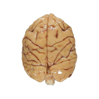
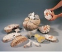
Preserved Sheep Brain, Dura Mater Removed, Plain, Pail
Quantity: 2
Sheep brains are the perfect specimen to study mammalian brain structure. A sheep brain is similar to a human brain and the structures are easily identified. Standard formalin sheep brains are shipped in Carosafe®, Carolina’s proprietary shipping and holding fluid. Carosafe® is an odorless fluid designed to minimize the unpleasant odor of formaldehyde. Brains are available in pails or in damp-packed, vacuum-sealed bags.
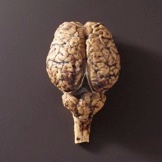
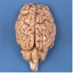
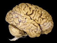
Horse Brain
Quantity: 1
This large, whole brain is an alternative to a calf brain. It features the hypophysis and some intact cranial nerves.

Pig Brain
Quantity: 1
Preserved whole pig brain.
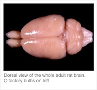
Rodent Brain
Quantity: 1
Preserved whole prairie vole and mouse brains are available.
Rhesus Macaque Brain
Quantity: 1
Preserved whole rhesus macaque brain is available.
Human Brain
Quantity: 2
Preserved whole and half (sagittal) human brains are available.
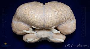
Dolphin Brain
Quantity: 2
Preserved whole dolphin brain.

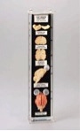
Comparative Brains Plastomount
Quantity: 1
Fish, amphibian, reptile, bird, and mammal brains mounted together. Size, 6 × 3”.

Sheep Brain Biosmount
Quantity: 1
Biosmount™ preparation. Dissected with external and internal views exposed. Two median sagittal sections and 2 transverse sections. Labeled. Size, 5 × 10 1/2”.
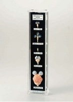
Brain Collection Museum Mount
Quantity: 1
Various Size and Shapes of Brain from Five Different Vertebrate Classes. Each brain includes the eyes. The five species represented are: cat, chicken, turtle or snake, frog, and perch. The cat cranial blood vessels are injected for better clarity. Each specimen is clearly labeled. Size: 4″W x 3″D x 15 3/4″H.
Cat Brain Anatomy Museum Mount
Quantity: 1
This mount is an excellent counterpart to the Brain Collection because it focuses on one species. Included in the collection is a whole brain with eyes, a median sagitally sectioned brain, plus cross and horizontal sections of the cerebrum and cerebellum. The cranial arteries of each specimen are injected with red pigment to highlight certain features. All specimens are mounted on a black plate and 12 structures are identified. Size: 3″W x 2″D x 15 1/4″H.
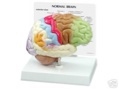
Half Brain-Sensory/Motor
Quantity: 1
Normal right half brain features: frontal, parietal, occipital and temporal lobes, cerebellum, interthalmic adhesion, corpus callosum, pons, midbrain-central peduncle, olfactory bulb, optic nerve, optic chiasm, mammillary body and medulla oblongata.
Nerve Synapse
Quantity: 1
Magnified many thousands of times, this depiction of a synapse is based upon electron microscopic interpretation. Sectioned open, the synaptic knob reveals its neurotubules, neurofilaments, synaptic vesicles, and the membranous ultrastructure of the postsynaptic apparatus.
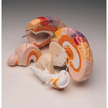
Giant Brain
Quantity: 1
Our largest brain model, this giant replica is 2.5 times life-size, and ideal for hands-on study in large groups. Highly detailed, all of the brain’s structures and the ventricles are visible through the median, frontal and horizontal sections. Sits on removable base.
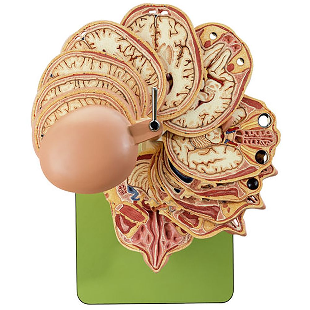
Horizontal Sectioned Model of Human Head
Quantity: 1
Life size. Model shows horizontal cross sections through the head and is oriented in the usual plane for CT and MR imaging. Includes 10 horizontal sections. Mounted on a base.
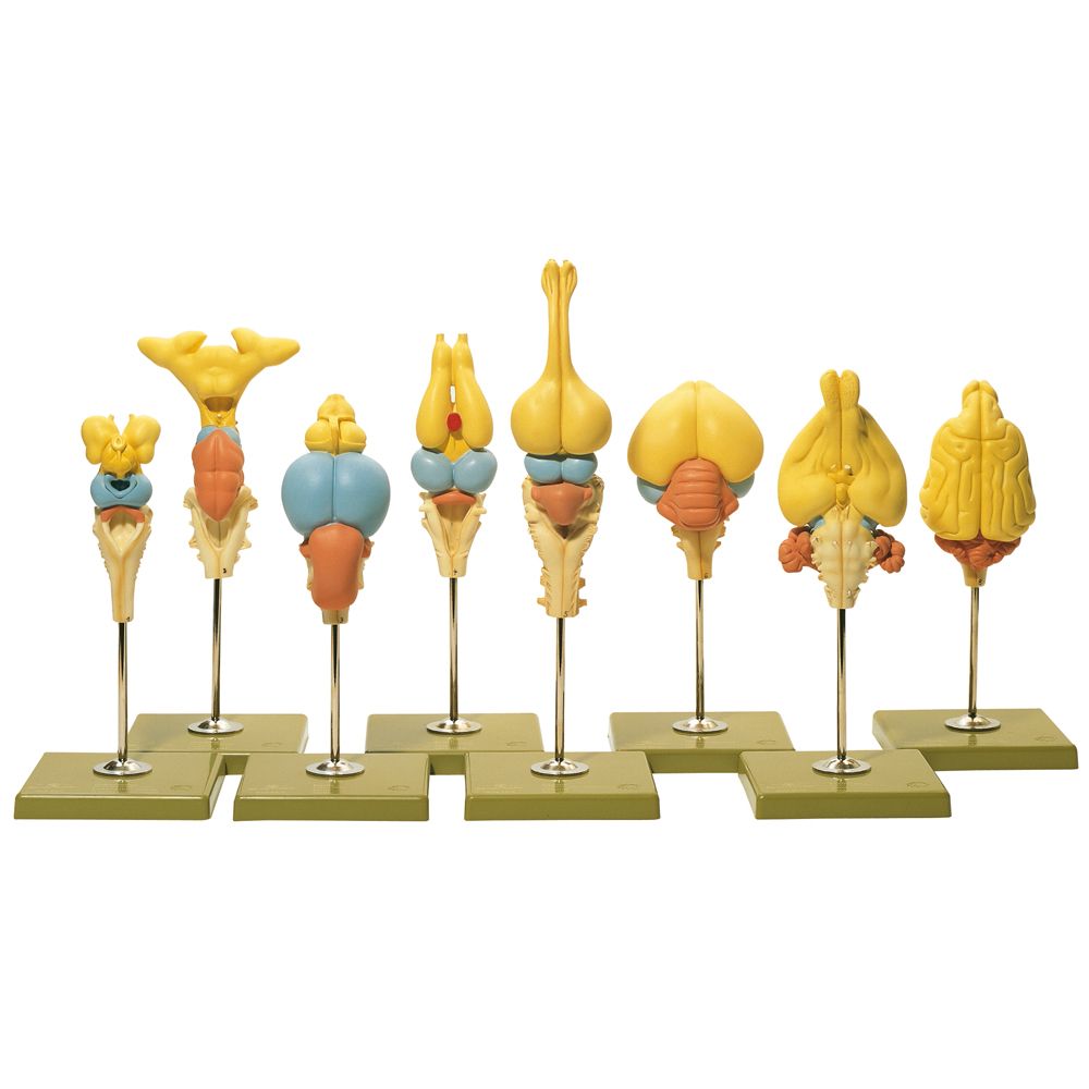
Vertebrate Models Brain Set
Quantity: 1 set of 8
Set of 8 individual, enlarged vertebrate brain models. Includes a lamprey, dogfish, trout, frog, alligator, dove, rabbit, and dog brain. Color-coded key included.
Functional Brain Model
Quantity: 1
2x life size. Depicting the brain of a right-handed person, contrasting colors and hand-lettered captions locate and identify motor and sensory functional centers, focus on the intellectual role of the dominant left brain, the creative role of the right, and call attention to the emotional, sexual, memory, and learning functions of the limbic system. Sensory regions highlighted include the separate reception and interpretive centers of the visual, acoustic, touch, taste, and olfactory senses.

Understanding Alzheimer’s Disease Chart
Quantity: 1
Defines Alzheimer’s disease and discusses the aging brain, dementia, and diagnosing Alzheimer’s disease. It lists risk factors, describes the three main stages of Alzheimer’s disease, and provides key management techniques for both patients and their families and caregivers. The chart illustrates and discusses the areas of the brain affected by the disease. Explains and shows a close up view of amyloid plaques (also called senile plaques). Describes and illustrates the following:
- Physical changes to the cerebral cortex
- Abnormal cellular structures involved (granulovacuolar degeneration and neurofibrillary tangles)
- Normal neuron and how Alzheimer’s disease destroys the way neurons “talk” to each other.
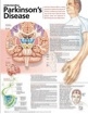
Understanding Parkinson’s Disease Chart
Quantity: 1
Defines Parkinson’s disease. Labeled illustrations include: Coronal section of the brain Lateral view of the brain Neurotransmitters- describes their role and the steps from a neuron receiving a message to post-message release of dopamine Shows a figure with a tremor impulse coming from the brain down the arm and explains the involvement of the brain with body movement Lists the causes and risk factors of Parkinson’s disease describes and illustrates common signs and symptoms lists related symptoms such as decreased sense of smell, depression, and sleep problems describes various management and treatment options such as medications, surgery, and therapy. Illustrates an electrode brain implant on a figure with a mask-like expression.
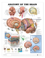
Anatomy of the Brain Anatomical Chart
Quantity: 1
Illustrations by renowned medical illustrator Keith Kasnot is one of our most popular charts. Beautiful, clear illustrations make the structures of the brain come alive. All illustrations are clearly labeled and vividly colored. Illustrations include:
- Central image showing major structures, cerebral hemispheres and key cranial nerves
- Arteries of the Brain (base and right side views)
- Venous Sinuses
- Lobes of the brain
- Cross-section of meninges & venous sinuses
- Typical nerve and glial cells
- Circulation of cerebrospinal fluid
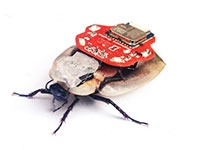
RoboRoach
Quantity: 1
With the RoboRoach you can briefly wirelessly control the left/right movement of a cockroach by microstimulation of the antenna nerves. The RoboRoach is a great way to learn about neural microstimulation, learning, and electronics! The RoboRoach “backpack” weighs 4.4 grams with the battery. Following a brief surgery you perform on the cockroach to attach the silver electrodes to the antenna, you can attach the backpack to the roach and control its movement for a few minutes before the cockroach adapts. When you return the cockroach to its cage for ~20 minutes, he “forgets” and the stimulation works again. **Must download 1x Free iOS or Android 4.3+ application for remote control and must order your own live roaches.
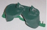
Prism Goggles
Quantity: 4
Perception goggles are plastic safety goggles with 45 degree prisms welded on to the goggle lens. The goggles are completely spray painted in dark paint, except for the angled face of the prisms. When students look through them, their vision is offset by about 30 degrees.
How to use: Students may play with the goggles one at a time or in groups. They may play with tennis balls or on the floor with coins or in front of the blackboard. What you can teach:
- You can demonstrate how quickly human brains can adapt to distorted incoming visual data.
- You can let your students have fun playing with their brains.
- You can demonstrate how the left motor strip controls the right side of the body and the right motor strip controls the left side of the body.
Concussion Goggles Kit
Quantity: 2
This kit is a hands-on awareness program that includes the specifically constructed Concussion Goggle™ used to simulate the potentially debilitating effects of Traumatic Brain Injury (TBI) caused by a bump, blow, or jolt to the head or body. Participants of the program experience the simulated TBI symptoms of dizziness, visual disconnect, disorientation, hesitation, apprehension, confusion, and lack of confidence. The program leaves each participant with an appreciation for their own susceptibility to TBI and what steps to take if someone sustains such an injury. Includes:
- 1x Concussion Goggle™
- 1x cloth protective bag
- 1x CD-Rom: QuickStart, classroom presentations, pre/post tests
- 1x roll of “walk the line” tape
- 1x triangle activity game
- 1x stress ball
- 1x Concussed™ card game
- 300x Action Steps pocket guides
- Germicidal Disposable Wipes
PBS NOVA: Secrets of the Mind
Quantity: 1
Ever been told “it’s all in your head”? Just how true is that statement? This provocative “Nova” documentary explores four puzzling case studies involving the mysteries of brain science. Join pioneering scientist V.S. Ramachandran as he investigates one person’s phantom limb pain, another’s messianic identity crisis, a young man’s certainty his parents are impostors, and the baffling conundrum of a blind man with the power of vision. 60 min. Soundtracks: English, DVS.
National Geographic: Brain Games
Quantity: 1
National Geographic’s groundbreaking three-part series provides a fascinating window into the inner workings of the brain as never before. Through interactive experiments and tricks, Brain Games reveals how our brains create the illusion of a seamless reality. As these revealing experiments provide a unique view into our brains, the world’s leading experts explain how and why these tests work. Brain Games explores cutting-edge science to examine real people with extraordinary brains, revealing new discoveries about attention, sensory perception and memory.